


How Eye Injuries Can Damage the Cornea
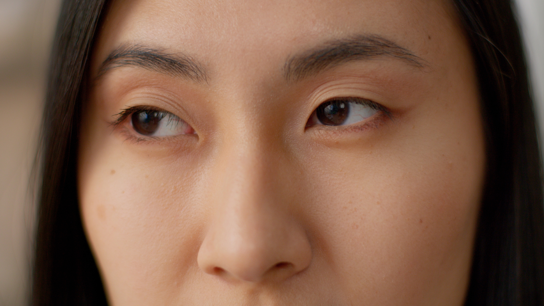



A stray grain of sand, a fingernail scratch or a ball striking the eye may seem like a minor incident. However, because the cornea forms the clear, protective front surface of the eye, even a small injury can cause considerable discomfort and interfere with vision. Eye injuries are particularly common during sports, outdoor activities and travel, when the eyes may be exposed to dust, debris, chemicals or intense ultraviolet light.
Corneal injuries range from surface scratches to deep wounds, burns and infections. Understanding the common types of corneal injury, how they affect vision and the available treatment options can help you recognise when specialist assessment is needed.
What Is the Cornea and Why Is It Vulnerable to Injury?
The cornea is the transparent, dome-shaped surface at the front of the eye. Because it is directly exposed to the environment, the cornea is vulnerable to dust, contact lenses, chemicals, flying objects and ultraviolet radiation.
Different corneal layers respond differently to injury. Superficial damage to the outer epithelium may heal quickly, while deeper stromal injuries are more likely to cause haze or scarring. Damage to the inner endothelial layer may lead to persistent swelling and reduced transparency.
Common Eye Injuries That Can Damage the Cornea
Corneal damage can result from a single accident or repeated irritation. Some injuries are immediately obvious, whereas others begin with mild discomfort and become more serious if an infection develops or a foreign body remains in the eye.
Corneal Abrasions
A corneal abrasion is a scratch or scrape on the outer surface of the cornea. Common causes include fingernails, makeup brushes, paper edges, tree branches, sand and particles trapped beneath the eyelid. Incorrect insertion or removal of a contact lens may also scratch the surface.
Abrasions can be intensely painful because the cornea contains many sensory nerve endings. Although many uncomplicated abrasions heal well, the eye should be assessed when pain is severe, vision changes or symptoms do not improve as expected.
Foreign Objects in the Eye
Dust, grit, glass, metal and plant material may land on or become embedded in the cornea. Blinking over a trapped particle can create repeated scratches, while organic material may carry microorganisms that increase the risk of infection.
An object that appears fixed in the eye should not be pulled out at home, as removal may worsen the wound or disturb tissue that is temporarily sealing it.
Blunt and Penetrating Eye Injuries
A ball, racket, elbow, fall or vehicle accident can compress the eye and damage several structures at once. Even when the outer appearance seems relatively normal, blunt force may affect the cornea, iris, lens, retina or optic nerve.
Chemical and Thermal Burns
Household cleaners, industrial chemicals, fertilisers, cement and laboratory substances can injure the cornea within a short period. Hot liquids, steam and flames can produce thermal injuries involving the cornea and eyelids.
Chemical exposure is an eye emergency. Immediate, continuous irrigation should begin before travelling for medical care.
Contact Lens-Related Injuries
Contact lenses can irritate or scratch the corneal surface when they are damaged, poorly fitted, worn for too long or handled incorrectly. More importantly, sleeping, showering or swimming in lenses, using water on lenses, or failing to clean and replace them as instructed can increase the risk of microbial keratitis.
Contact lens wearers who develop pain, eye redness, discharge, light sensitivity or blurred vision should remove their lenses and seek prompt professional advice.
Ultraviolet Eye Injuries
Intense ultraviolet exposure can injure the corneal epithelium, producing a condition known as photokeratitis. It may follow welding without suitable protection or prolonged exposure to sunlight reflected from snow, water or sand.
How Corneal Injuries Can Affect Your Vision
Corneal injuries can disrupt the eye’s protective surface or alter its clarity. Central, deep, contaminated or slow-healing wounds carry a greater risk of lasting visual effects.
Infection and Corneal Ulcers
Damage to the corneal surface can allow microorganisms to enter, causing keratitis or a corneal ulcer. Severe ulcers may lead to thinning, scarring or perforation. Risk can be influenced by contact lens wear, contaminated water, plant material and delayed treatment.
Corneal Scarring and Clouding
Deeper wounds may heal with haze or scar tissue. A central scar can blur vision, reduce contrast and cause glare. Mild haze may improve during healing, while persistent clouding may require further treatment.
Permanent Vision Loss in Severe Cases
Severe burns, penetrating wounds and uncontrolled infections can cause lasting visual impairment, particularly when other eye structures are also damaged. Early treatment can control infection and preserve tissue, although full visual recovery cannot always be guaranteed.
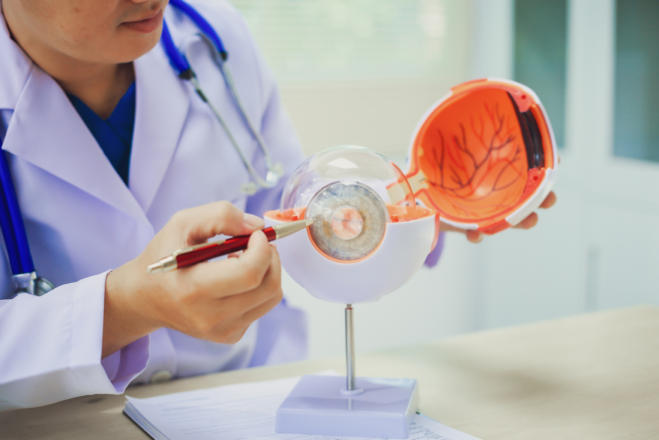
How Are Corneal Injuries Treated?
Treatment depends on the cause, depth, location and cleanliness of the wound, as well as whether infection or damage to other parts of the eye is present.
Treatment for Corneal Abrasions
Management of an uncomplicated abrasion may include lubricating drops or ointment, pain relief and prescribed medication when clinically appropriate. Follow-up may be recommended for larger abrasions, contact lens wearers or injuries that are not improving.
Treatment for Corneal Infections and Ulcers
Suspected microbial keratitis requires prompt treatment. Depending on its appearance and severity, the cornea may be sampled to help identify the responsible organism.
Antibacterial, antifungal or other antimicrobial eye drops can then be selected and adjusted according to the clinical response and laboratory findings.
Repair of Deep Corneal Wounds
A deep or full-thickness laceration may require an operation to restore the integrity of the eye. Fine sutures can be used to align the wound, while tissue adhesive and a protective contact lens may be suitable for selected small leaks.
Specialised Corneal Injury Assessment at Eye & Cornea Surgeons
Although some corneal injuries heal with appropriate care, deeper wounds, infections and chemical burns can lead to scarring or lasting changes in vision. Persistent pain, redness, light sensitivity or blurred vision should therefore be assessed promptly to determine the extent of the damage and reduce the risk of complications.
At Eye & Cornea Surgeons, patients have access to an experienced team of ophthalmologists. The team includes Prof Donald Tan Tiang Hwee, Dr Chan Wing Kwong and Dr Anna Tan Wee Tien, who have extensive experience in diagnosing and treating corneal conditions, including complex cases that may require corneal surgery or transplantation.
If you are experiencing persistent symptoms following an eye injury, book an appointment with our clinic.
FAQs About Corneal Injuries
Can corneal abrasion symptoms return after the eye has healed?

What type of eye protection can help prevent corneal injuries?
What should I do if eye-injury symptoms develop while travelling?
Meet Our Ophthalmologists in Singapore
Prof Donald Tan Tiang Hwee
MBBS(S'pore), FRCS(Glas), FRCS(Edin), FRCOphth(UK), FAMS
Ophthalmologist
Partner and Senior Consultant
Prof Donald Tan Tiang Hwee is a partner and senior consultant at Eye & Retina Surgeons, where he leads the Eye & Cornea Surgeons division. An internationally recognised authority in corneal disease and transplantation, he has pioneered advances in corneal surgery and helped develop modern corneal transplant techniques used around the world. With decades of clinical, research and academic leadership, Prof Tan specialises in the management of complex corneal and ocular surface disorders.
Dr Chan Wing Kwong
MBBS(S'pore), MMed(Ophth), FRCS(Edin), FRCOphth, FAMS
Ophthalmologist
Partner and Senior Consultant
Dr Chan Wing Kwong is a partner and senior consultant at Eye & Retina Surgeons and a visiting senior consultant at the Singapore National Eye Centre. A fellowship-trained corneal and refractive surgeon, he has extensive experience in the diagnosis and treatment of corneal diseases, cataracts and refractive conditions. Widely recognised for his expertise in laser vision correction and corneal surgery, Dr Chan combines decades of clinical experience with a commitment to delivering personalised eye care.
Dr Anna Tan Wee Tien
MBBS (S'pore), MMed (Ophth), MRCSEd, FAMS
Ophthalmologist
Partner and Senior Consultant
Dr Anna Tan Wee Tien is a partner and senior consultant at Eye & Retina Surgeons and a fellowship-trained corneal and refractive surgeon. Formerly Head of Cornea and External Eye Diseases at the National University Hospital, she has extensive experience managing corneal disorders and performing corneal transplantation, cataract surgery and laser vision correction procedures. Dr Tan is dedicated to providing comprehensive, patient-centred care tailored to each individual's eye health needs.